41 neuron model with labels
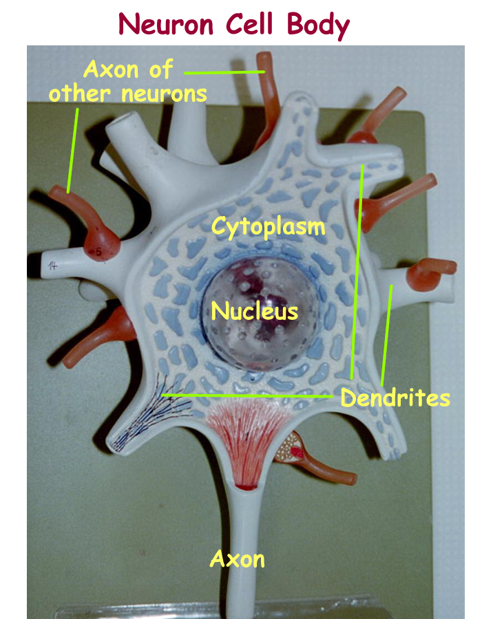
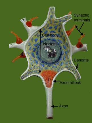
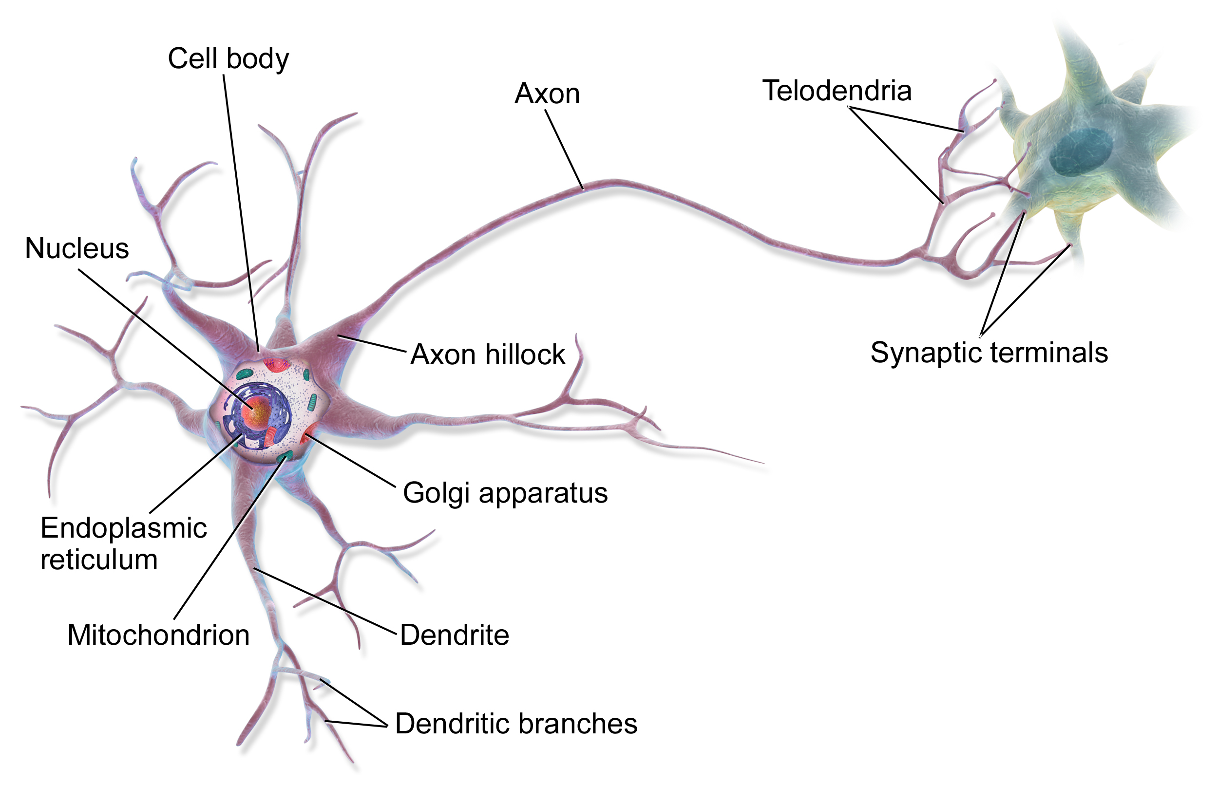
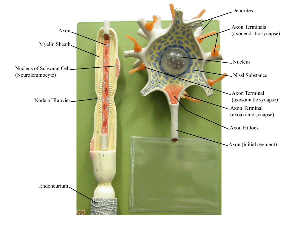
12.2 Nervous Tissue - Anatomy and Physiology 2e | OpenStax Figure 12.8 Parts of a Neuron The major parts of the neuron are labeled on a multipolar neuron from the CNS. Where the axon emerges from the cell body, ... Some cutting edge research suggests that certain neurons in the CNS do not conform to the standard model of "one, and only one" axon. Some sources describe a fourth type of neuron ... Neuron Models - San Diego Mesa College Neuron Models. Click on a photo for a larger view of the model. Click on Label for the labeled model. Back to Nervous System. Neuron. Cell Body & Dendrites. Axon.
Amazon.com: Neuron Model Jackson Global JS00134 Neuron Model - 2 Parts | 2500X Enlarged | Labeled Diagram Included. $86.52 $ 86. 52. FREE delivery Thu, Apr 27 . Only 2 left in stock - order soon. ... Amazon's Choice for neuron model. nerdbugs Neuron (Brain Cell) Plush - Neuron My Mind! -Brain Cell Plush Organ/ Get Well Gift/ Health Education Toy/ Neuroscience or ...

Neuron model with labels
12.2 Nervous Tissue - Anatomy & Physiology Figure 12.2.2 - Parts of a Multipolar Neuron: The major parts of the neuron are labeled on a multipolar neuron from the CNS. External Website. Visit this site (link not working as of 10/20/2021) to learn about how nervous tissue is composed of neurons and glial cells. Neurons are dynamic cells with the ability to make a vast number of ... Neuron Diagram & Types | Ask A Biologist - Arizona State University Unipolar neurons are also known as sensory neurons. They have one axon and one dendrite branching off in opposite directions from the cell body. These cells pass signals from the outside of your body, such as touch, along to the central nervous system. Bipolar neurons have one axon and only one dendrite branch. Neuroscience for Kids - Models - University of Washington This giant model of a neuron illustrates the properties of chemical transmissionand the action potential. Cut two to three foot lengths of rope to use as dendrites. foot piece of rope will be turned into the axon. The cell body and synaptic terminal of the neuron can be plastic containers. Drill holes in
Neuron model with labels. Neuron Model - YouTube For pictures of this model with answer keys to help you study, visit: ... Labelled Diagram Of Motor Neuron - schematron.org Description, AO1: The Structure and Function of Sensory, Relay and Motor Neurons The nervous system is composed of specialised cells called neurons. Worksheet with diagrams of sensory and motor neurones to label and a table with summary of functions. Sensory neurons bring signals into the CNS, and motor neurons carry _Image modified from ... Overview of neuron structure and function - Khan Academy Neurons are the basic functional units of the nervous system, and they generate electrical signals called action potentials, which allow them to quickly transmit information over long distances. Glia are also essential to nervous system function, but they work mostly by supporting the neurons. A new color-coded map of the C. elegans nervous system The approach is fundamentally different from the previously described Brainbow method used to label individual neurons in different model organisms, including C. elegans.Whereas Brainbow is a ...
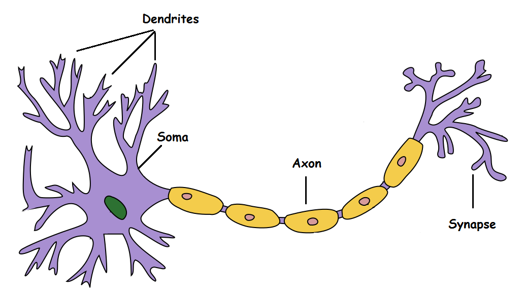
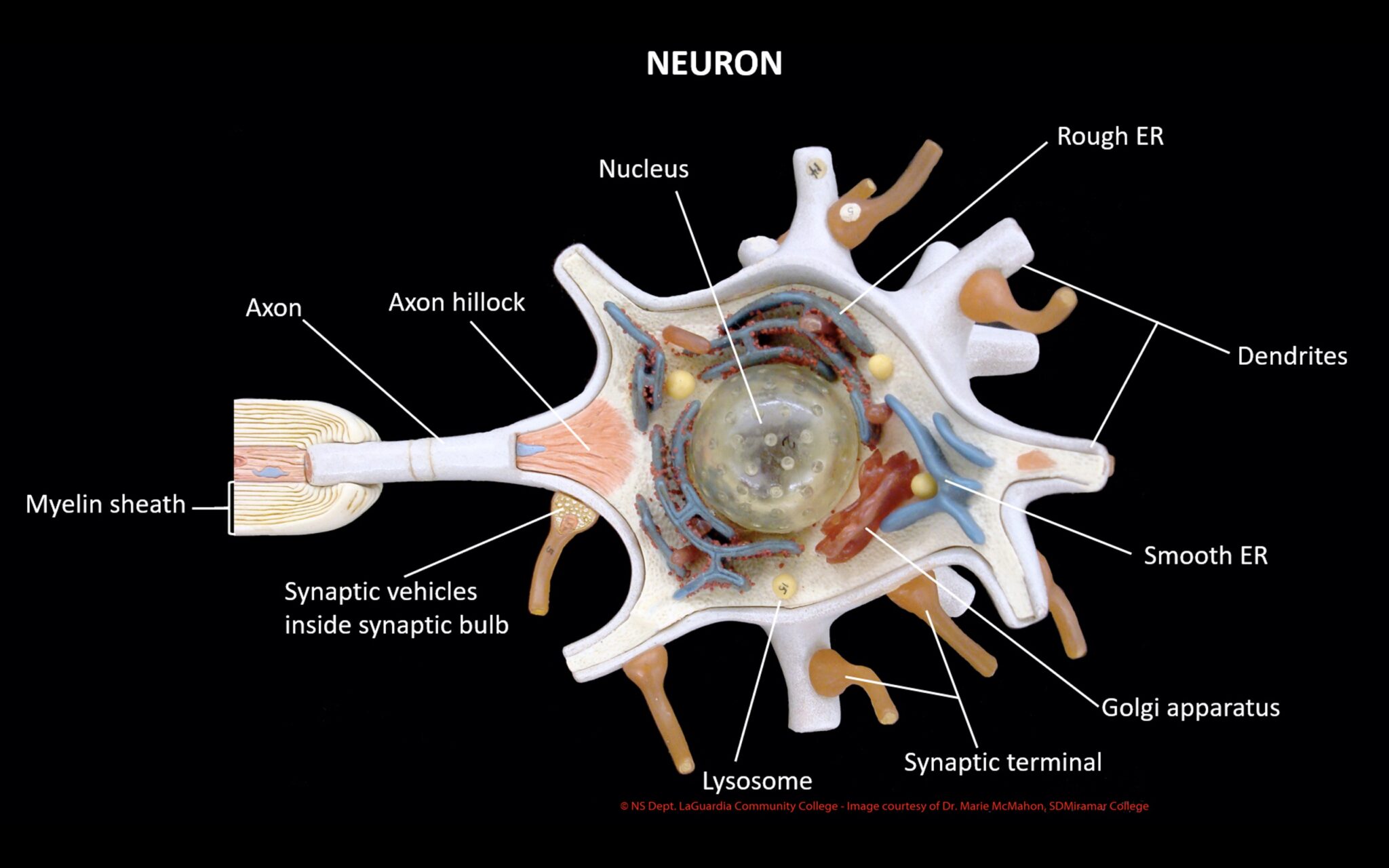
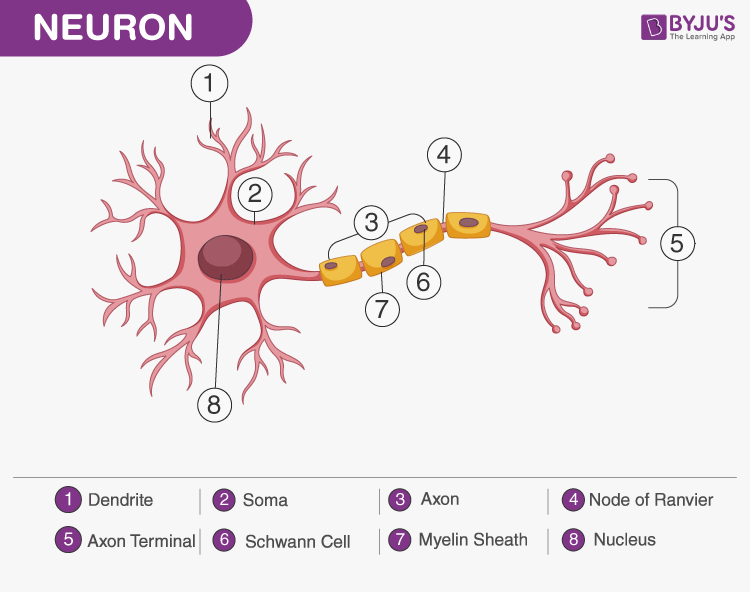
A Labelled Diagram Of Neuron with Detailed Explanations - BYJU'S Diagram Of Neuron with Labels Here is the description of human neuron along with the diagram of the neuron and their parts. The neuron is a specialized and individual cell, which is also known as the nerve cell. A group of neurons forms a nerve. Neuron Model Anatomy - YouTube neuron model for anatomy and physiology Diagram Quiz on Neuron Structure and Function (Labeling Quiz) 1. Identify the cell type in the above figure Liver Cell Cardiac Cell Nerve cell Skin cell 2. In the figure, labeled '1' receives impulses from adjacent neuron. It is called the Dendron Dendrite Axon Axonite 3. In the figure, labeled '2' is the short filaments from the cell body that carries impulses from dendrites to the cell body which is the Hodgkin-Huxley model - Wikipedia The Hodgkin-Huxley model, or conductance-based model, is a mathematical model that describes how action potentials in neurons are initiated and propagated. It is a set of nonlinear differential equations that approximates the electrical characteristics of excitable cells such as neurons and muscle cells.It is a continuous-time dynamical system.. Alan Hodgkin and Andrew Huxley described the ...
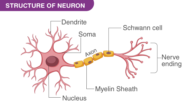
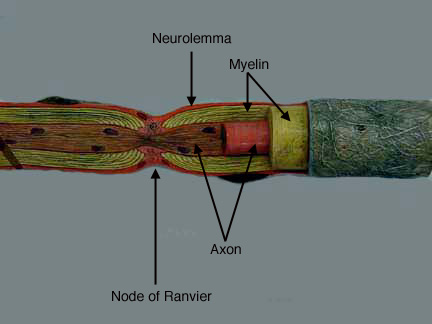
The synapse (article) | Human biology | Khan Academy Neurons communicate with one another at junctions called synapses. At a synapse, one neuron sends a message to a target neuron—another cell. Most synapses are chemical; these synapses communicate using chemical messengers. Other synapses are electrical; in these synapses, ions flow directly between cells. Neuron Model Labeling Diagram | Quizlet Neuron Model Labeling + − Flashcards Learn Test Match Created by lindsey_lasater0 Terms in this set (7) Term Node of Ravier Definition periodic gap in the insulating sheath (myelin) on the axon of certain neurons that serves to facilitate the rapid conduction of nerve impulses. Location Neuron a specialized cell transmitting nerve impulses. Term Parts of a Neuron and Their Functions with Labelled Diagram - Science Facts All neurons have three main parts: 1) dendrites , 2) cell body or soma, and 3) axons. Besides the three major parts, there is the presence of axon terminal and synapse at the end of the neuron. 1) Dendrites They are specialized extensions that resemble the branch of a tree. Label a Neuron Model Quiz - PurposeGames.com Label a Neuron Model by mp2000 361 plays 11 questions ~30 sec English 11p More 0 too few (you: not rated) Tries Unlimited [?] Last Played February 22, 2022 - 12:00 am There is a printable worksheet available for download here so you can take the quiz with pen and paper. From the quiz author Practice labeling structures of a neuron model. Remaining
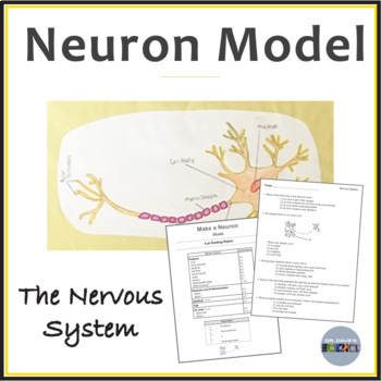
Cards ,Crafts ,Kids Projects: How to make a Neuron Model
Neuron Labeling Using Best Practices (8 Tips) - label template A neuron chart is a graphical representation of brain cells, making labeling neurons easy and precise. A neuron chart can be made with paper or online and customized to fit your needs. To create a neuron chart, start by clustering all the cells in your image into groups according to their function.
Biology 2404 A&P Basics
Spinal cord: Anatomy, structure, tracts and function | Kenhub Anatomy. The spinal cord is part of the central nervous system (CNS). It is situated inside the vertebral canal of the vertebral column. During development, there's a disproportion between spinal cord growth and vertebral column growth. The spinal cord finishes growing at the age of 4, while the vertebral column finishes growing at age 14-18.

608 Neuron Labeled Images, Stock Photos & Vectors | Shutterstock
What Is a Neuron? Diagrams, Types, Function, and More - Healthline Neurons, also known as nerve cells, send and receive signals from your brain. While neurons have a lot in common with other types of cells, they're structurally and functionally unique....

What Is a Neuron? Diagrams, Types, Function, and More
Identify Neuron Model Quiz - purposegames.com This online quiz is called Identify Neuron Model. It was created by member Katie Cavins and has 8 questions. ... Label Parts of the Brain. Medicine. English. Creator. ninalahoti. Quiz Type. Image Quiz. Value. 12 points. Likes. 57. Played. 182,183 times. Printable Worksheet. Play Now. Add to playlist.

Overview of neuron structure and function (article) | Khan ...
Single-neuron mechanisms of neural adaptation in the human temporal ... The dominating psychological model for semantic-relatedness priming is the spreading activation model 10. In our data we do not find evidence in favor of this model as neither iEEG nor single-unit ...
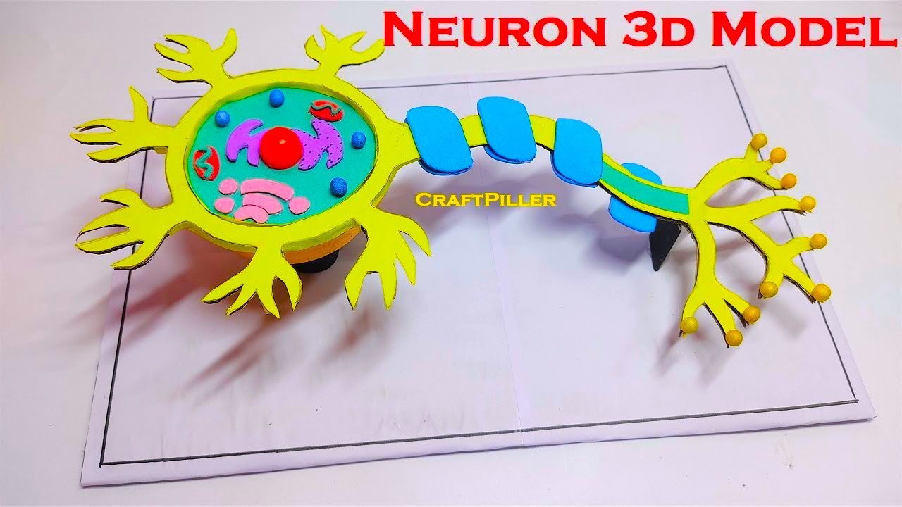
neuron model making(3D) | biology model diy | craftpiller | still model
Keras: show loss for each label in a multi-label regression Suppose you have a Keras model with n neurons as the output, where each neuron is associated to a regression variable (e.g. speed of a car, height of a car, ...), as in the following code snippet: # ... I'd like to get a history object that contains the loss for each of the n labels, i.e. {loss: {'speed': loss_value_speed, 'height': loss_value ...
Neuron Models
A Guide to Understand Neuron with Neuron Diagram To properly understand the coordination between the brain and the body, the students must learn about the neurons. They can use neuron-labeled diagrams while learning the complex structure of neurons. Creating a neuron-labeled image by hand can be difficult. The students must use the EdrawMax Online tool to make a high-quality neuron diagram . 2.
McCulloch-Pitts Neuron — Mankind's First Mathematical Model ...
Neuroscience for Kids - Models - University of Washington This giant model of a neuron illustrates the properties of chemical transmissionand the action potential. Cut two to three foot lengths of rope to use as dendrites. foot piece of rope will be turned into the axon. The cell body and synaptic terminal of the neuron can be plastic containers. Drill holes in

neuron structure model making project DIY | howtofunda | science project | still model
Neuron Diagram & Types | Ask A Biologist - Arizona State University Unipolar neurons are also known as sensory neurons. They have one axon and one dendrite branching off in opposite directions from the cell body. These cells pass signals from the outside of your body, such as touch, along to the central nervous system. Bipolar neurons have one axon and only one dendrite branch.
What Is a Neuron? - Definition, Structure, Parts and Function
12.2 Nervous Tissue - Anatomy & Physiology Figure 12.2.2 - Parts of a Multipolar Neuron: The major parts of the neuron are labeled on a multipolar neuron from the CNS. External Website. Visit this site (link not working as of 10/20/2021) to learn about how nervous tissue is composed of neurons and glial cells. Neurons are dynamic cells with the ability to make a vast number of ...
Solved] Activity 1: Identifying parts of a neuron 1. You will ...
Neuron - Wikipedia
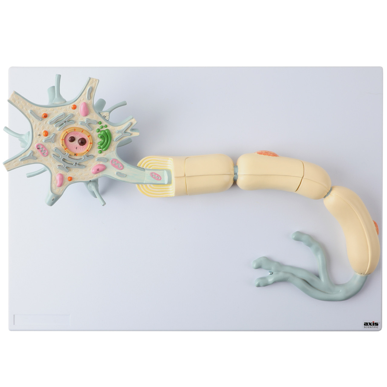
Axis Scientific Two-Part Neuron Model with Dendrite and Axon

Nervous and Spinal Cord Models
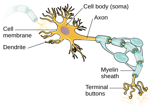
Neurons | Introduction to Psychology
Model of a Neuron Nervous System Activity Human Body ...
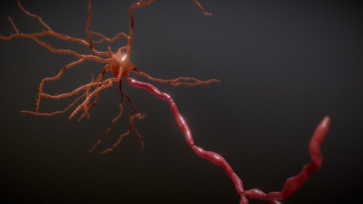
Neuron 3D models - Sketchfab
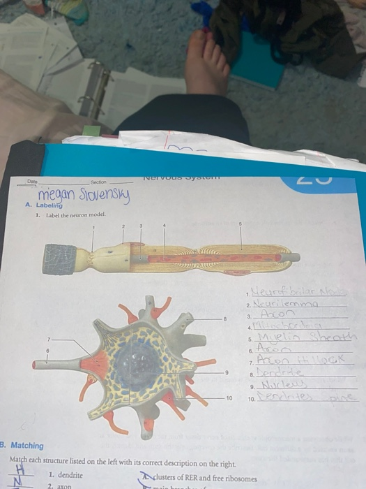
Solved Dan Nervous system Section megan Slovensky A. | Chegg.com
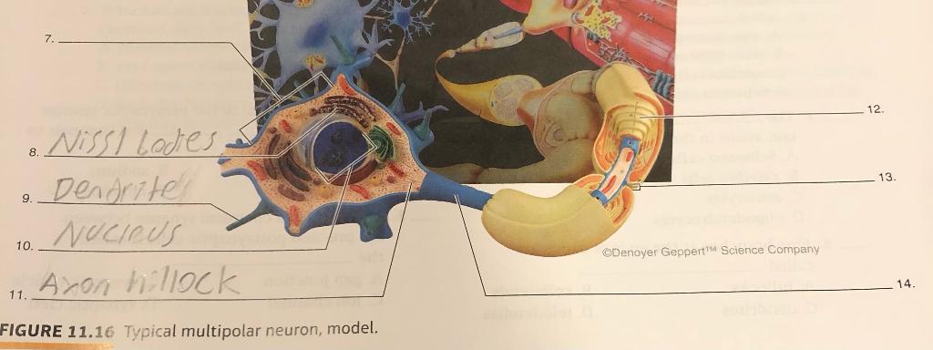
Solved Well I want to know if I labeled the ones correctly ...

Neuron model
neuron

Model Neuron, Neuron Cell Body Nerve Fiber Structure And ...
![Biological neuron model taken from [13] | Download Scientific ...](https://www.researchgate.net/profile/Mohd-Hafizul-Afifi-Abdullah/publication/330926855/figure/fig1/AS:777213658013700@1562313584976/Biological-neuron-model-taken-from-13_Q640.jpg)
Biological neuron model taken from [13] | Download Scientific ...

Organization of Cell Types (Section 1, Chapter 8 ...

File:Neuron-no labels.png - Wikimedia Commons

Motor Neuron, Detailed And Accurate, Labeled Stock Clipart ...

model of multipolar neuron Diagram | Quizlet

Cermin neuron Sistem saraf Sel Saraf, yang lain, bermacam ...

Diagram Berlabel Neuron Ilustrasi Stok - Unduh Gambar ...
SCB209 - Lab1 - Natural Sciences Open Educational Resources
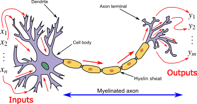
Mimicking Biological Neural Network | Artificial Neurons and ...

Pin on School
A Labelled Diagram Of Neuron with Detailed Explanations

Biological neuron model - Wikipedia
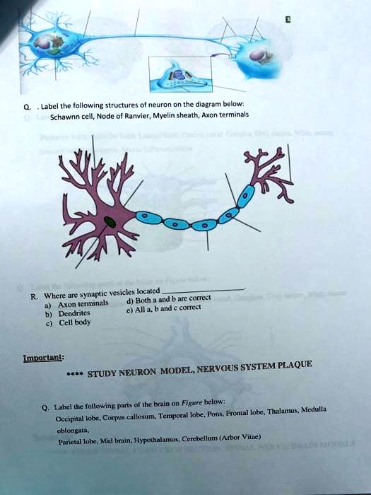
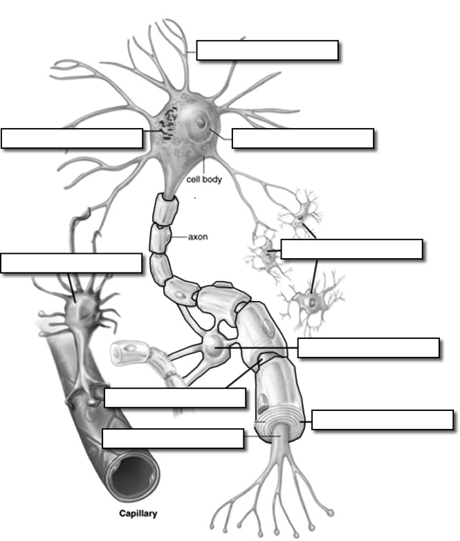
SOLVED: Q..Label the following structures of neuron on the ...

Neuron Model labeling Diagram | Quizlet

Neuron Model – Human Body Help
Neuron Label

Neuron Model Diagram | Quizlet

608 Neuron Labeled Images, Stock Photos & Vectors | Shutterstock
Neuron Models
Post a Comment for "41 neuron model with labels"